


Ultrasound and Doppler
Ultrasound scanning is a versatile diagnostic tool that can assist in detecting and evaluating a wide range of diseases across various medical fields.
About Ultrasound and Doppler
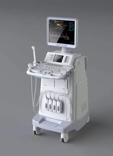
At Xpert Clinic, our team of highly skilled ultrasound and Doppler specialists uses state-of-the-art equipment to provide accurate and reliable diagnostics. We take pride in being among the pioneers to introduce 4D ultrasound scanning in India in 2004.
3D-4D Ultrasound
Xpert Clinic is a leader in 3D-4D ultrasound services.
Xpert Clinic offers the expertise to perform a wide range of routine and advanced studies, including FNACs, ultrasound-guided biopsies, and Fibroscan with Shearwave Elastography, across our centers in a patient-friendly and comfortable environment.
Ultrasound is a safe and non-invasive imaging technique that uses high-frequency sound waves, which are completely harmless to the body. It is highly versatile and can aid in diagnosing a variety of conditions. Ultrasound can be used to examine numerous areas of the body, including the abdomen and pelvis, musculoskeletal system, breast, male reproductive system, kidneys, thyroid and salivary glands, gallbladder, pancreas, liver, neck, and the developing fetus. When performed by skilled professionals, ultrasound is a reliable tool that can address most diagnostic questions effectively.
Please Note:
As per the PC-PNDT Act, performing a pregnancy ultrasound without a valid prescription, along with the doctor’s DMC/HMC/MCI registration number, phone number, and other required details, is strictly prohibited.
General, Musculoskeletal, and Vascular Ultrasound
Ultrasound scans are simple and safe procedures that create images of the body’s internal structures without using radiation. They can be used to examine various parts of the body, including:
Blood vessels
Pelvis
Heart
Musculoskeletal system
Abdomen
Other soft tissue organs
Pregnancy Ultrasound
Ultrasound is a highly valuable diagnostic tool, especially during pregnancy, as it is completely safe for both mother and baby, using no ionizing radiation. Some important uses of ultrasound during pregnancy include:
Determining the gestational age and accurately estimating your due date
Identifying the number of babies
Detecting any early pregnancy bleeding
Checking the position of the placenta
Monitoring the growth and overall well-being of the baby
Assessing the baby’s anatomy and screening for potential abnormalities*
*Please note that while ultrasound is highly useful for detecting many birth abnormalities, not all conditions can be reliably diagnosed. Results should always be interpreted within the limitations of the test.
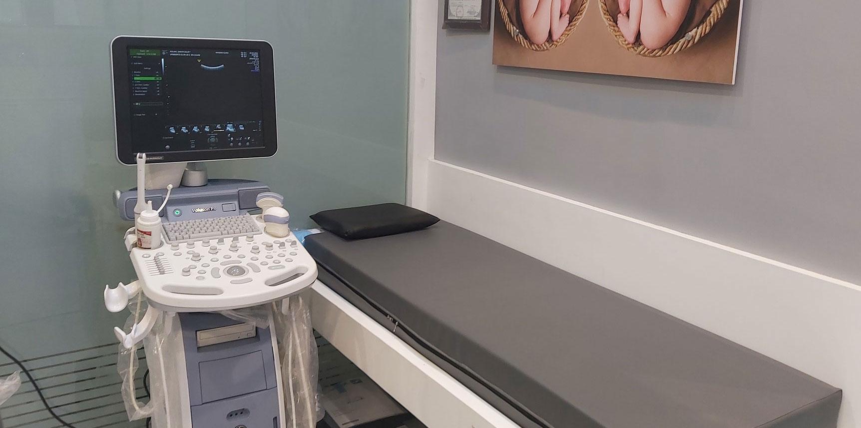
Shear Wave Fibrosis Scan
Shear Wave Elastography at Xpert Clinic, performed on advanced machines like LOGIQ E9 and Aplio i800, is a highly effective, non-invasive method for accurately measuring liver tissue stiffness.
Fibroscan (Liver Elastography)
Fibroscan is a non-invasive test that evaluates the health of your liver. Using ultrasound technology, it measures the level of fibrosis or scarring caused by various liver conditions. This test is particularly helpful for patients with fatty liver to identify those with progressive fibrosis, as well as for assessing fibrosis and cirrhosis of the liver from any underlying cause.
Liver Fibrosis Scan Using Shear Wave Elastography with Ultrasound
Quantitative Measurement of Tissue Stiffness: Provides precise stiffness values.
Shear Wave Speed (m/s) & Young’s Modulus (kPa): Measures the elasticity of liver tissue.
2D Color-Coded Visualization: Offers a clear, visual representation of tissue stiffness.
B-Mode Ultrasound Guidance: Enhances imaging with:
Instant assessment of spatial stiffness, aiding in detection and characterization of focal lesions
Overlay of shear wave stiffness maps on B-mode images for better diagnostic interpretation
Accurate placement of Regions of Interest (ROI) for precise stiffness measurements
Localized Sampling: Measures tissue stiffness at specific points within the ROI, unlike conventional Fibroscan which provides only an average stiffness blindly.
Follow-Up Assessment: Enables comparison of fibrosis levels at the same liver site over time, which was not possible with older techniques.
Colour Doppler
Cutting Edge Technologies
Colour Doppler is a medical imaging technique that visualizes blood flow by adding color to ultrasound images, allowing doctors to clearly observe the direction and speed of blood movement within the body. A duplex ultrasound, on the other hand, evaluates how blood flows through arteries and veins by combining traditional ultrasound with Doppler technology. While regular ultrasound uses sound waves to create images of blood vessels, Doppler analyzes the reflection of sound waves from moving objects, such as blood, providing detailed information about circulation.
Advanced Colour Doppler at Xperts Imaging
There are different types of duplex ultrasound exams. Some include:
- Abdominal arterial and venous duplex ultrasound evaluates the blood vessels and blood flow in the abdominal region.
- Carotid duplex ultrasound assesses the carotid arteries in the neck.
- Duplex ultrasound of the extremities examines blood flow in the arms and legs.
- Renal duplex ultrasound evaluates the kidneys and their blood vessels.
Why It Is Done
- Detect blood clots and identify blocked or narrowed blood vessels in various parts of the body, particularly in the neck, arms, and legs.
- Assess abnormal veins that may cause varicose veins or other related issues.
- Evaluate blood flow to a transplanted kidney or liver.
- Monitor blood circulation after blood vessel surgery.
Frequently Asked Questions (FAQ’S)
Why is an Ultrasound Doppler Test Done?
At Xpert Clinic, an Ultrasound Doppler test is performed to assess blood flow in veins and arteries. Using advanced sound wave technology, it measures the direction and speed of blood circulation, helping doctors detect issues such as restricted blood flow, narrowed arteries, and blood clots.
How is a Doppler Ultrasound different from a regular ultrasound?
A Doppler Ultrasound focuses on measuring blood movement within vessels by detecting changes in sound wave frequency caused by moving blood cells, while a regular ultrasound captures static images of organs, tissues, and internal structures. Unlike a standard ultrasound, a Doppler scan provides real-time visualization of blood flow, offering deeper insight into circulatory health.
How much time does an Ultrasound Doppler test take?
The duration of an Ultrasound Doppler test varies based on the type of scan being performed. Typically, it takes around 20 to 50 minutes to complete.
When will I get my Ultrasound Doppler test results?
Is an Ultrasound Doppler test able to detect blood clots?
Yes, an Ultrasound Doppler test can effectively detect blood clots. It is commonly used to diagnose conditions such as deep vein thrombosis. The test is non-invasive, provides real-time results, and works by evaluating blood flow to identify any abnormalities.
Is a contrast dye needed for a Doppler Ultrasound?
A Doppler Ultrasound uses sound waves to evaluate blood flow and detect issues like narrowed arteries or blood clots without the need for contrast dye. Therefore, contrast is typically not required for this test.
How reliable is an Ultrasound Doppler test?
The accuracy of an Ultrasound Doppler test depends on the quality of the equipment, the patient’s condition, and the expertise of the technician. Generally, it delivers reliable results in detecting issues such as narrowed arteries, venous insufficiency, and blood clots.
Are there any special instructions to follow after an Ultrasound Doppler test?
You can resume your normal activities immediately, as no special instructions are required after a Doppler Ultrasound. However, if a follow-up is advised, be sure to consult your healthcare provider.
Will the Ultrasound Doppler scan cause any pain or discomfort?
A Doppler Ultrasound scan is generally painless. You may only feel mild pressure when the technician moves the probe over the area being examined.